 Download this patient information sheet
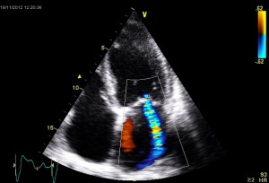
An echo (echocardiogram or cardiac ultrasound) examines structure of the heart non-invasively. It takes 20-30 minutes to perform and provides very useful information.
An echo gives important information about how the heart looks and functions. This is often key to how heart problems are treated and followed. It is one of the commonest heart tests performed.
You will be asked to change into a hospital gown. Three electrocardiogram (ECG) dots will be attached to your chest to record your heart tracing during the examination. The echo technician or cardiologist will ask you to lie on the bed and turn onto your left side. Ultrasound gel will be placed on your chest and the ultrasound probe will be placed onto your chest directly to obtain accurate pictures of your heart.
No.
Aside of some usually minor discomfort associated with the probe being placed on the chest there is no known risk. Similar ultrasound is used to examine babies inside the mother’s womb.
Suite 209, Level 2, Concord Hospital Medical Centre,
Hospital Rd,
Concord NSW 2139